An echocardiogram diagnoses an enlarged heart and other cardiac abnormalities that cause chest pain and shortness of breath. At Florida Heart, Vein and Vascular Institute, with offices in Zephyrhills, Riverview, Plant City, Wesley Chapel and Lakeland, Florida, echocardiogram testing is available to assess the health and function of your heart valves and other structures. The practice’s skilled cardiologists use the results of your echocardiogram to create an effective treatment plan for underlying heart issues. Call the Florida Heart, Vein and Vascular Institute office nearest you to learn more about the benefits of an echocardiogram, or book an appointment online today.
We offer comprehensive diagnostic and preventive heart and vein care, ensuring every patient receives accurate evaluation and effective treatment.
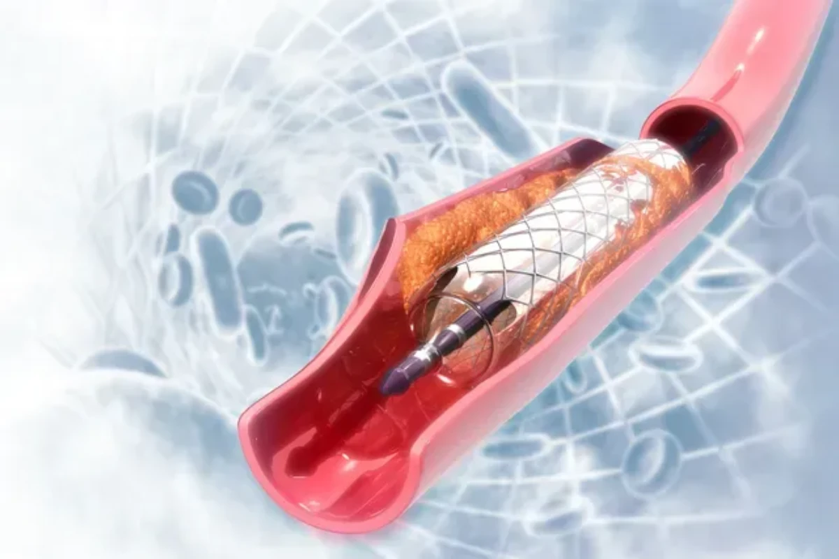
We specialize in minimally invasive procedures that open blocked arteries and restore proper blood flow, improving heart function and patient outcomes.
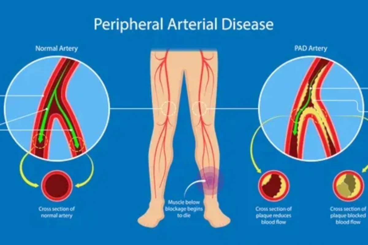
Our experts diagnose and treat narrowed or blocked vessels outside the heart, enhancing circulation and reducing pain in the legs and extremities.
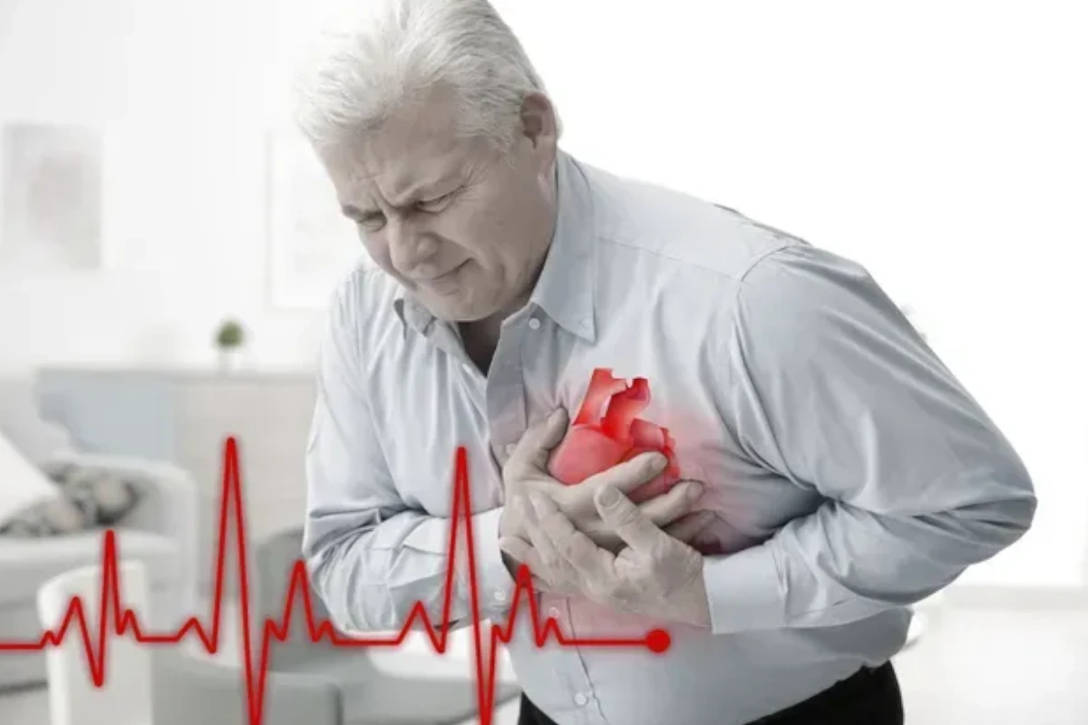
We provide advanced care to manage plaque buildup in coronary arteries, improving oxygen flow to the heart and preventing serious cardiac events.
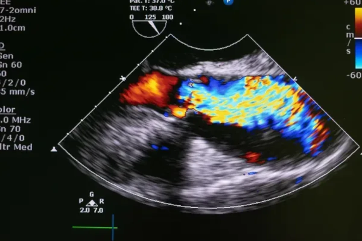
Our specialists assess and treat valve disorders to restore normal heart rhythm and ensure efficient blood flow throughout the cardiovascular system.
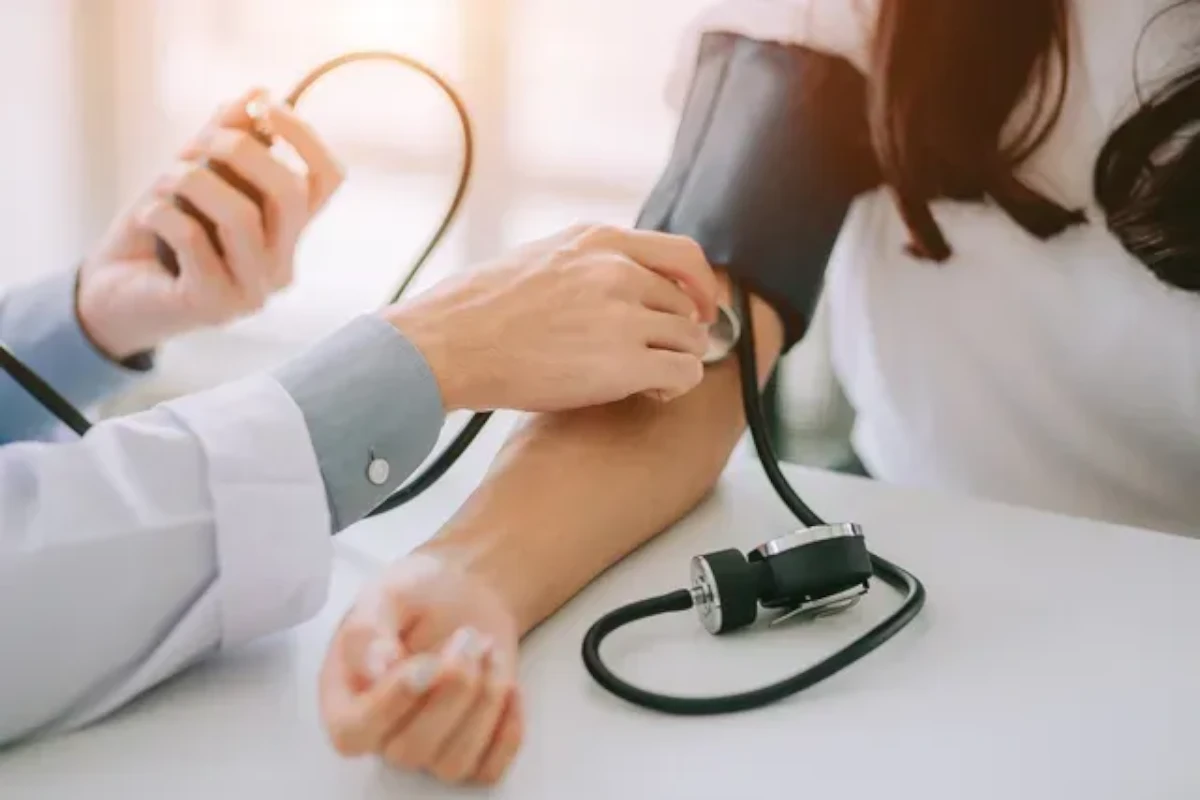
We offer personalized management for high blood pressure through medication, diet, and lifestyle adjustments to protect your heart and arteries.
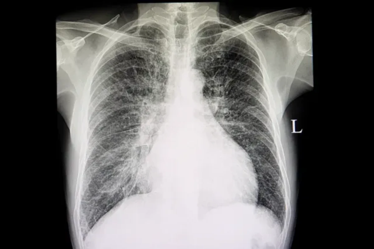
Comprehensive treatment plans help strengthen heart function, reduce symptoms, and improve quality of life for patients with heart failure.
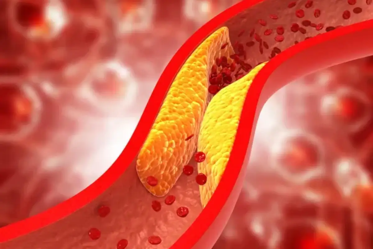
We focus on preventing heart disease through cholesterol management programs that combine medication, nutrition counseling, and lifestyle coaching.
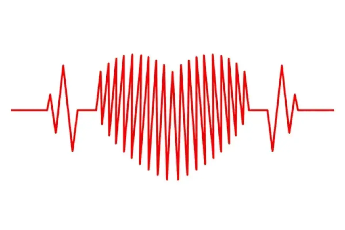
Our team evaluates irregular or rapid heartbeats with advanced diagnostic tools to identify causes and develop effective treatment strategies.
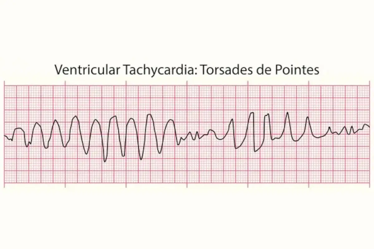
We diagnose and manage heart rhythm abnormalities using state-of-the-art monitoring and therapies that help maintain a steady, healthy heartbeat.
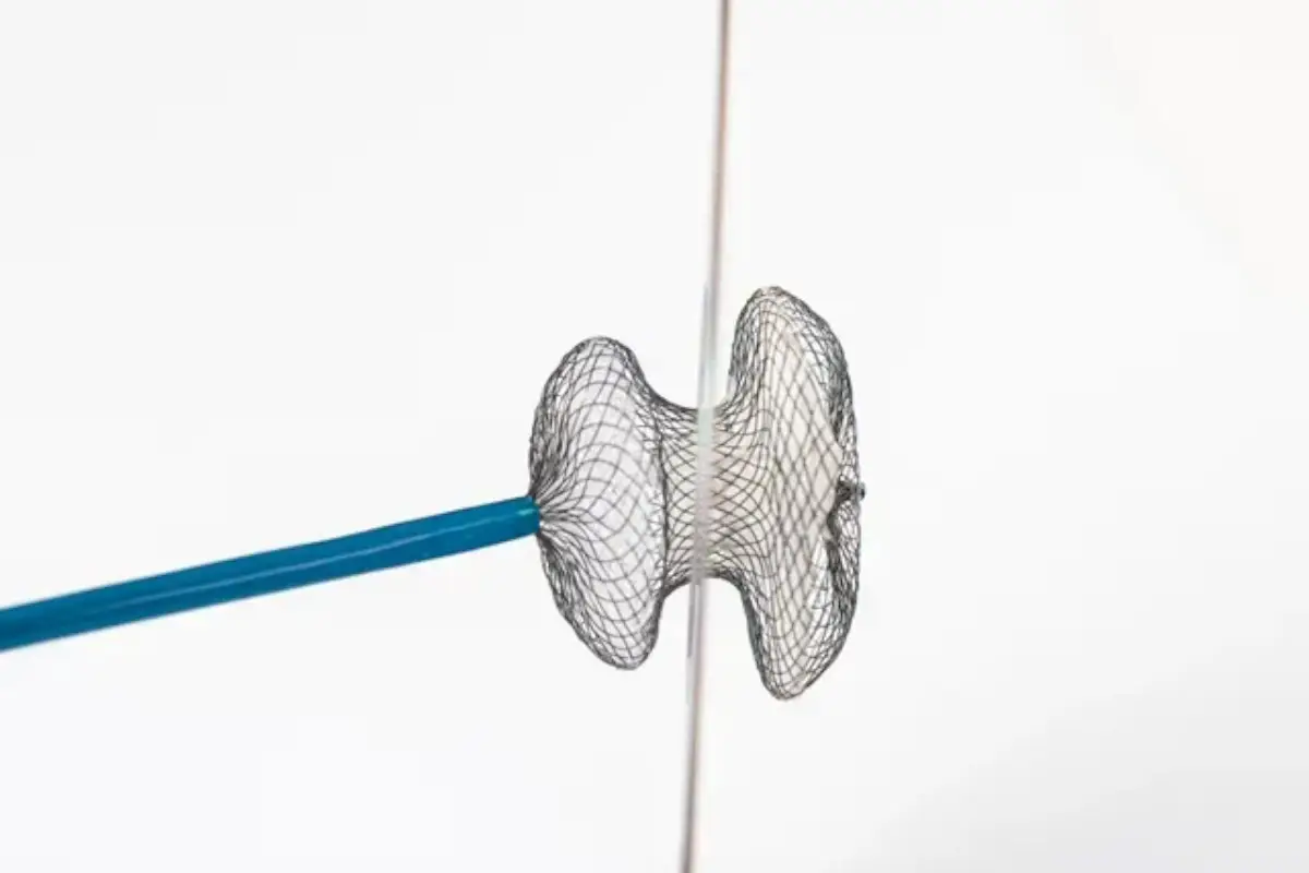
We offer cutting-edge care for atrial fibrillation, using medication and minimally invasive procedures to restore normal rhythm and prevent stroke.

Our specialists investigate fainting episodes to determine underlying cardiovascular causes and provide treatment to prevent future occurrences.
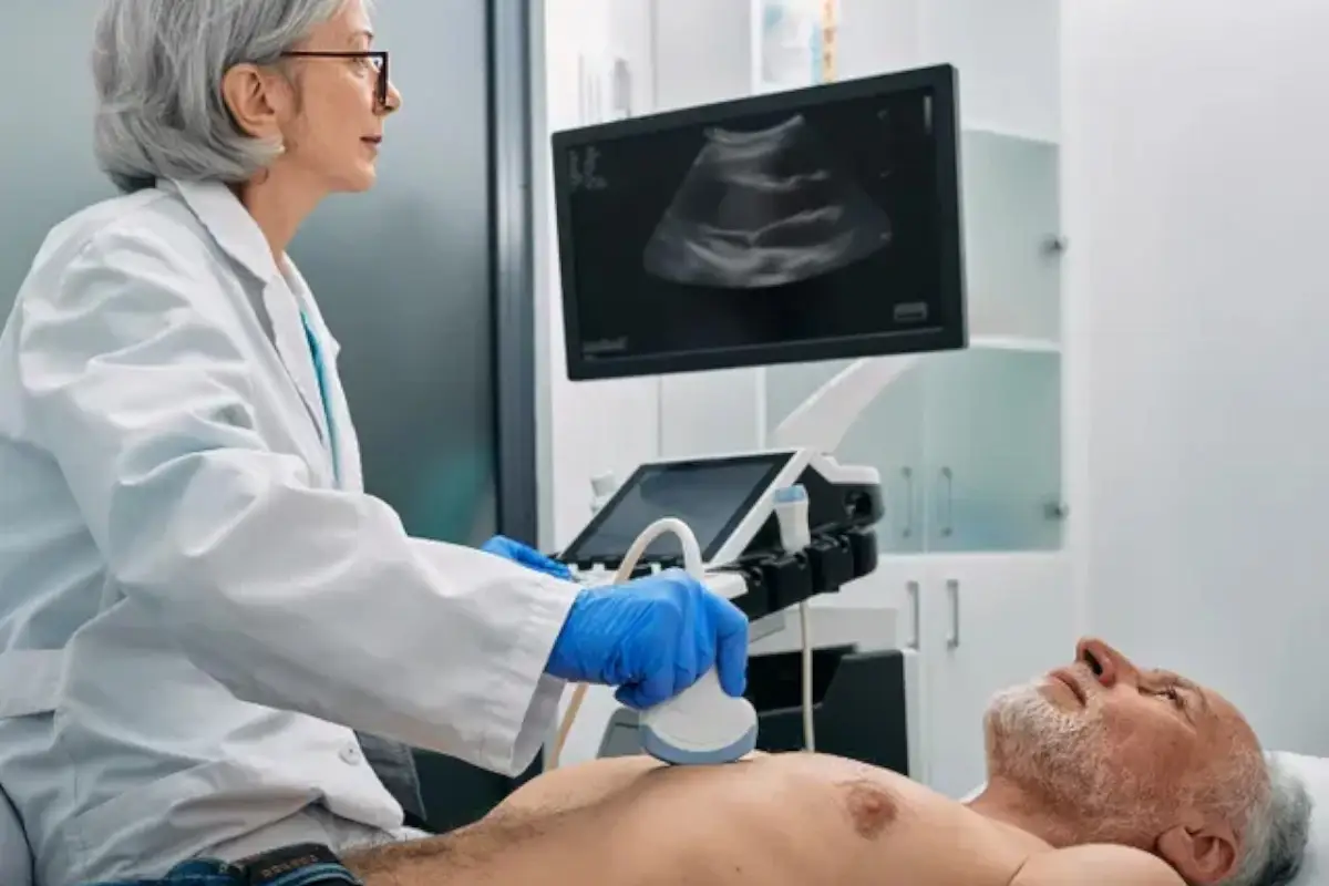
This non-invasive ultrasound test gives detailed images of your heart’s structure and function, helping us assess heart health and detect conditions early.
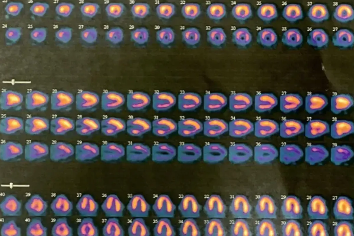
We use advanced imaging to evaluate blood flow to the heart muscle, identifying areas of reduced circulation or damage with high precision.
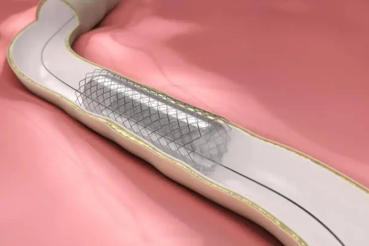
We diagnose and treat varicose veins, deep vein issues, and other venous conditions to restore comfort, improve circulation, and prevent complications.
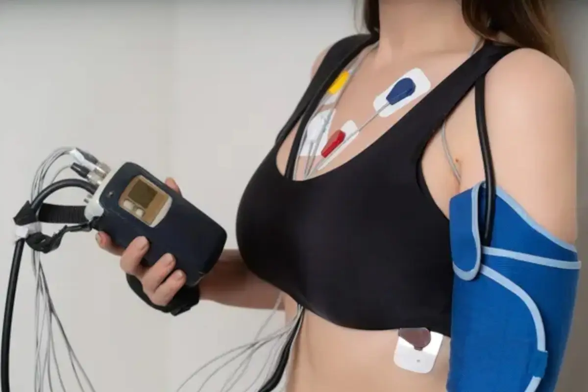
A portable Holter device records your heart’s activity over 24–48 hours, allowing accurate detection of rhythm irregularities in real-life settings.
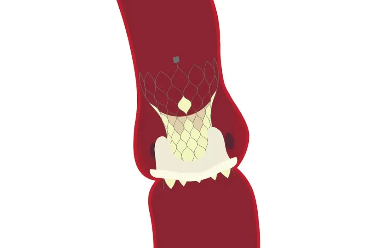
TAVR offers a minimally invasive alternative to open-heart surgery, replacing damaged valves to improve blood flow and overall heart performance.
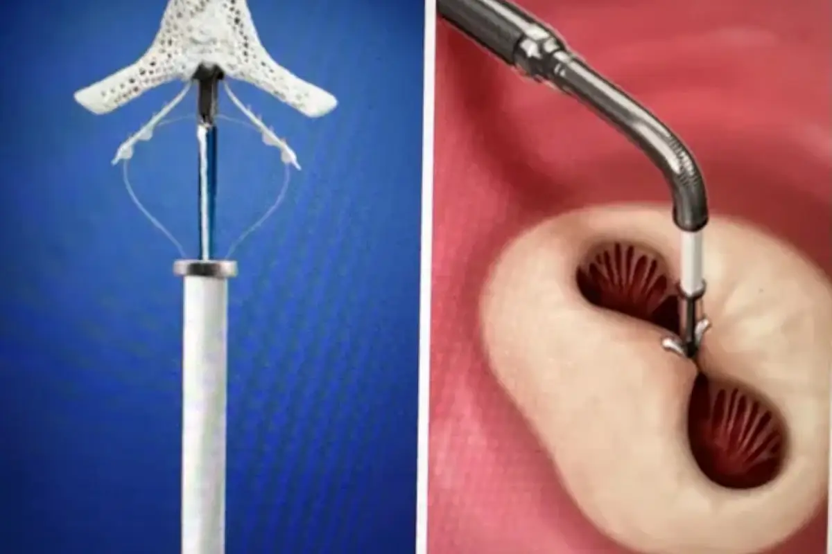
We perform MitraClip therapy to treat mitral valve regurgitation, improving heart efficiency and reducing symptoms without the need for surgery.
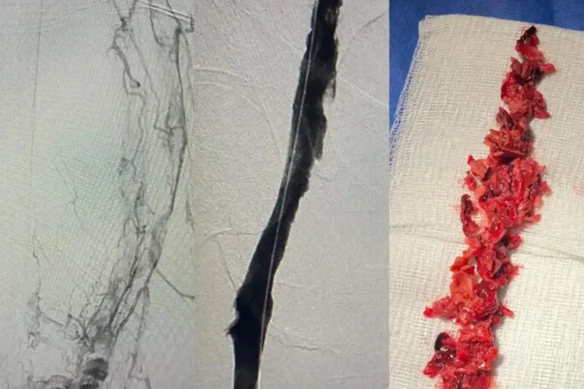
Our specialists use advanced, minimally invasive techniques to dissolve clots, relieve pain, and prevent long-term vein damage from DVT.
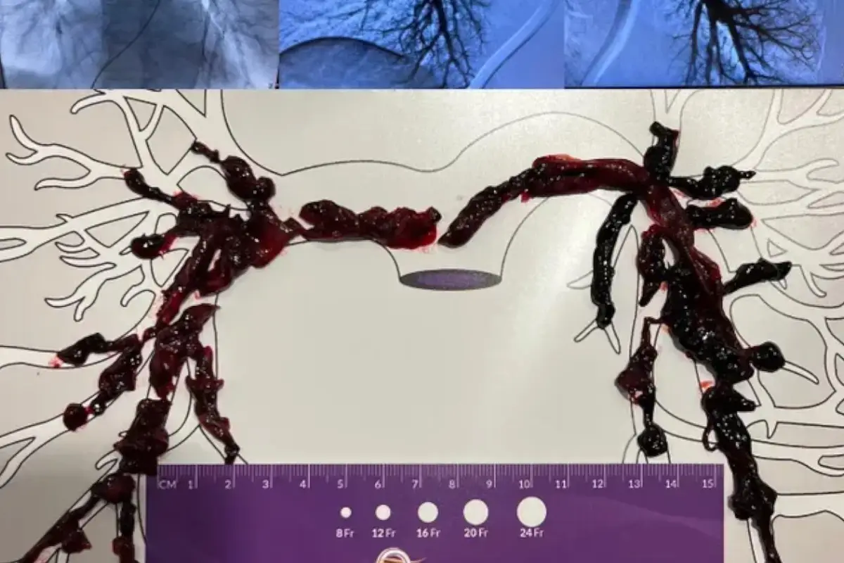
We offer rapid diagnosis and targeted treatments to remove or dissolve clots in the lungs, restoring breathing and reducing cardiovascular risk.
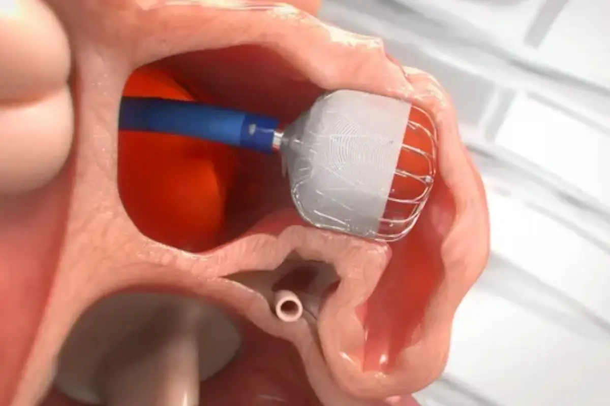
This innovative procedure reduces stroke risk in atrial fibrillation patients by closing the left atrial appendage and eliminating long-term anticoagulants.
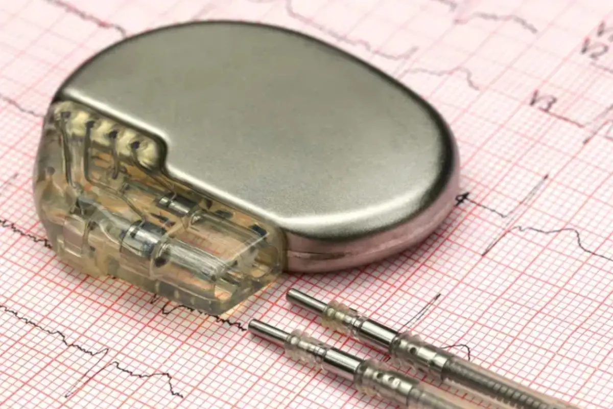
We implant pacemakers to regulate slow or irregular heartbeats, restoring proper rhythm and improving daily function and energy levels.
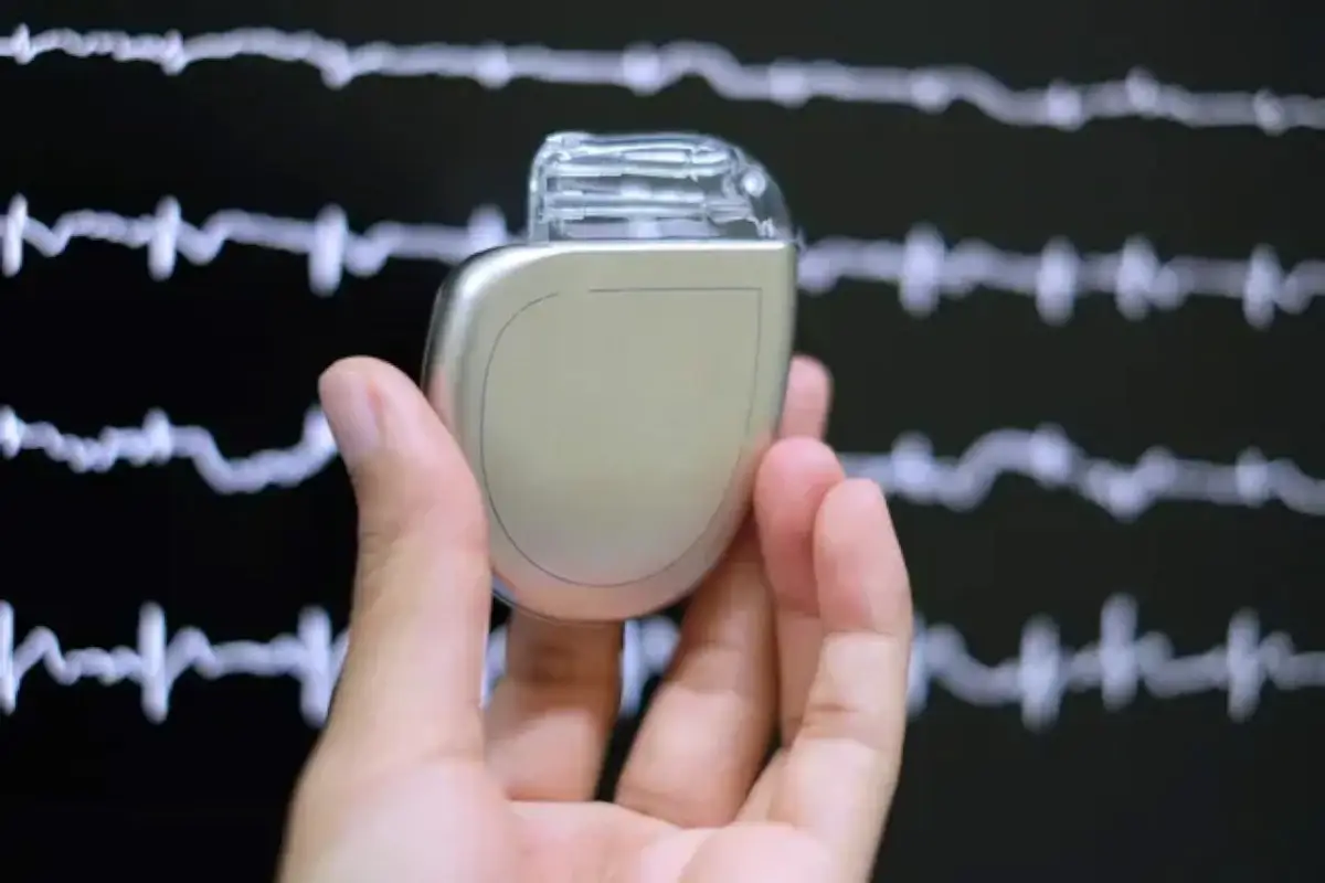
Our ICD implantation helps prevent sudden cardiac arrest by detecting dangerous rhythms and automatically delivering corrective electrical pulses.
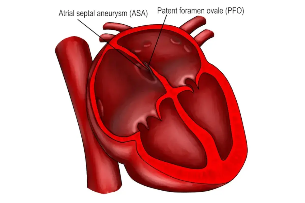
We close the small opening between heart chambers using a catheter-based device, reducing the risk of stroke and improving overall circulation.